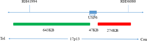
USP6(17p13) gene break apart probe reagent
10 Tests/box
This kit performs fluorescence in situ hybridization staining on the basis of conventional staining, and provides auxiliary information for diagnosis for physicians. The test results are for clinical reference only and should not be used as the sole basis for clinical diagnosis.Clinicians should make comprehensive judgments on the test results based on factors such as the patient’s condition, drug indications,treatment response and other laboratory test indicators.
The kit is based on fluorescence in situ hybridization technology. A nucleic acid probe islabeled with fluorescein. The target gene is detected with homologous complementary to the nucleic acid probe used. Both after denaturation, annealing and renaturation, the hybrid of the target gene and the nucleic acid probe can be formed, and the qualitative, quantitative or relative positioning analysis of the gene to be measured under the microscope can be performed by the fluorescence detection system.
The kit consists of USP6 dual-color probes, as shown in Table 1.
| Component name | Specifications | Quantity | Main components |
|---|---|---|---|
| USP6 dual color probe | 100μL/Tube | 1 | USP6 orange probe ; USP6 green probe |
Keep sealed away from light at -20oC±5oC, and the validity period is 12 months. After the cover is opened, it can be sealed and stored in 2~8°C away from light within 24 hours. After the cover is opened, it should be sealed and stored in -20±5°C away from light for a long time. Transport with temperature below 0°C.
Fluorescence microscopy imaging systems, including fluorescence microscopy and filter sets suitable for DAPI (367/452), Green (495/517),and Orange (547/565).
1. Applicable specimen types: Paraffin-embedded specimens from surgical excision or biopsy.
2. The tissue should be fixed with 4% neutral formaldehyde solution within 1 hour after isolation. After tissue fixation, it is routinely dehydrated and embedded in paraffin.
3. Paraffin section thickness could affect the experimental results. The recommended slice thickness is 4~5μm.
4. Paraffin-embedded tissue samples from breast cancer should be selected from representative tumor tissue wax blocks and confirmed by HE staining.
5. It is recommended to select paraffin-embedded tissue specimens within 5 years preservation period.
Instructions
1. Hybridization pretreatment
Baking: Slides heating at 80oC for 30min or 65oC for 2h or overnight.
Dewaxing: According to the customer laboratory protocol (Commonly with Xylene for 15min).
Hydration: Take out the slides and put them respectively into 100%, 85% and 70% EtOH at room temperature for 3 minutes each.
Take out the slides, and immerse them in deionized water for 3 minutes. Remove the excess of water on the slides by air-drying.
Permeation: Immerse the slides in deionized water at 100oC and boil continuously for 20-40 minutes (Conventional 20min). Remove the excess of water on the slides by air-drying.
Digestion: Protease enzymic digestion at 37°C for 10-40 minutes. Mix the protease work buffer (50mmol HCl) and the 10x protease solution (Pepsin concentration 5%) in a proportion of 9:1 to prepare the enzymatic digestion solution.
Washing: Wash with 2xSSC at room temperature for 5 minutes.
Dehydration: Take out the slides and dehydrate in 70%, 85%, and 100% gradient ethanol at room temperature for 2 minutes each time.Remove the excess of EtOH solution on the slides by air-drying.
2. Denaturation and Hybridization
The following operations should be performed in a darkroom.
3. Washing
The following operations should be performed in a darkroom.
4. Counterstaining
The following operations should be performed in a darkroom.
Dip 10μL of DAPI counterstain into the hybridization area of the glass slides, immediately cover with a lid and place in dark for 10-20min,then use the appropriate filter to observe the sections under the fluorescence microscope.
5. FISH results observation
Place the counterstained film under the fluorescence microscope, and first put it under the low-power objective lens (10x) confirm the cell area under the microscope; Go to 40x under the objective lens, find a position where the cells are evenly distributed; Then in the high-power objective (100x) the FISH results of nuclei were observed.

Negative: 2 fusion

Positive: 1 orange 1 green 1 fusion
The results of this kit will be affected by various factors of the sample itself, as well as by the limitations of hybridization temperature and time, operating environment, and limitations of current molecular biology techniques, which may lead to erroneous results.Users must be aware of potential errors and limitations of accuracy that may exist in the detection process.
1. Fluorescence signal intensity: after the probe is effectively hybridized with the karyotype sample, it shall send out a fluorescence signal that can be recognized by the naked eye under the fluorescence microscope.
2. Sensitivity: detect karyotype samples and analyze the chromosomes of 50 cells in metaphase division phase. At least 98 chromosomes 17 show an orange fluorescence signal and a green fluorescence signal.
3. Specificity: the chromosomes of 50 cells in metaphase division phase were analyzed. At least 98 chromosomes 17 showed a specific orange fluorescence signal in the target region and a specific green fluorescence signal in the centromere region.
1. Please read this manual carefully before testing. The testing personnel shall receive professional technical training, and the signal counting personnel must be able to observe and distinguish orange and green signals.
2. When testing clinical samples, when it is difficult to count the hybridization signal and the samples are not enough to repeat the retest,the test will not provide any test results. If the amount of cells is insufficient for analysis, the test will not provide test results.
3. Formamide and DAPI counterstaining agent used in this experiment have potential toxicity or carcinogenicity. It is necessary to operate in the fume hood and wear masks and gloves to avoid direct contact.
4. The results of this kit will be affected by various factors of the sample itself, but also limited by enzyme digestion time, hybridization temperature and time, operating environment and the limitations of current molecular biology technology, which may lead to wrong interpretation results. Users must understand the potential errors and accuracy limitations that may exist in the detection process.
5. All chemicals are potentially dangerous. Avoid direct contact. Used kits are clinical waste and should be properly disposed off.
[1]. K. E. Olsen et al. Amplification of HER2 and TOP2A and deletion ofTOP2A genes in breast cancer investigated by new FISH probes,ActaOncologica 43(2004)
[2]. K. V. Nielsen et al. The value of TOP2A gene copy number variation asa biomarker in breast cancer: Update of DBCG trial89D,ActaOncologica, 2008; 47: 725734
[3]. Zaczek, Markiewicz, Seroczynska et al. Prognostic Significance of TOP2A Gene Dosage in HER-2-NegativeBreast Cancer, The Oncologist 2012;17:1246–1255
[4]. Järvinen TA, Tanner M, Bärlund M, et al. Characterization of topoisomerase II alpha gene amplification and deletion in breast cancer.Genes Chromosomes Cancer. 1999 Oct; 26(2):142-50.
[5]. Li Xuyuan, Li Yubing, Lin Ying Cheng breast cancer TOP2A gene and anthracycline efficacy, [J] International Journal of oncology 2011.2.38 (2).
V1. 0: Approval date: November 4, 2019.
V1. 2: Revision date: December 7, 2021.