Product Cat. No.: GBS-053

For Research Use Only.

FUS gene break apart probe reagent Instructions Manual

Product Name

FUS gene break apart probe reagent.

Package Specifications

10 Tests/box.

Product Introduction

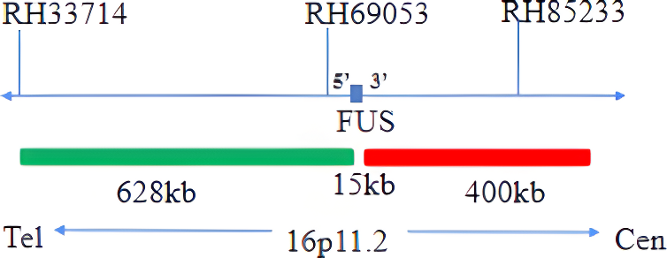
This kit uses Orange fluorescein and Green fluorescein to label FUS. FUS probe can be bound to the target detection site by in situ hybridization.

Product Main Components

The kit consists of FUS dual color probe as shown in Table 1.

Table 1: Kit composition

Component name Specifications Quantity Main components
FUS dual color probe 100μL/Tube 1 FUS orange probe ; FUS green probe

Storage conditions & Validity

The kit should be transported below 0°C. Keep sealed away from light at -20oC±5oC. The product is valid for 12 months. Avoid unnecessary repeated freezing and thawing that should not exceed 10 times. After opening, within 24 hours for short-term preservation, keep sealed at 2-8oC in dark. For long-term preservation after opening, keep the lid sealed at -20oC±5oC away from light.

Applicable Instruments

Fluorescence microscopy imaging systems, including fluorescence microscopy and filter sets suitable for DAPI (367/452), Green (495/517), and Orange (547/565).

Sample requirements

  1. Applicable specimen types: Paraffin-embedded specimens for surgical resection or biopsy.
  2. Tissue should be fixed with 4% neutral formaldehyde fixation solution within 1 hour after ex vivo, and the tissue should be fixed by conventional dehydration and paraffin embedding.

Instructions

  1. Pre-hybridization or Pretreatment
    Baking: Slides heating at 80oC for 30min or 65oC for 2h or overnight.
    Dewaxing: According to the customer laboratory protocol (Commonly with Xylene for 15min).
    Hydration: Take out the slides and put them respectively into 100%, 85% and 70% EtOH at room temperature for 3 minutes each.
    Take out the slides, and immerse them in deionized water for 3 minutes. Remove the excess of water on the slides by air-drying.
    Permeation: Immerse the slides in deionized water at 100oC and boil continuously for 20-40 minutes (Conventional 20min). Remove the excess of water on the slides by air-drying.
    Digestion: Protease enzymic digestion at 37°C for 10-40 minutes. Mix the protease work buffer (50mmol HCl) and the 10x protease solution (Pepsin concentration 5%) in a proportion of 9:1 to prepare the enzymatic digestion solution.
    Washing: Wash with 2xSSC at room temperature for 5 minutes.
    Dehydration: Take out the slides and dehydrate in 70%, 85%, and 100% gradient ethanol at room temperature for 2 minutes each time. Remove the excess of EtOH solution on the slides by air-drying.
  2. Denaturation and Hybridization
    The following operations need to be carried out in the darkroom.

    1. Take out the probe, leave it at room temperature for 5min, turn it upside down with force, mix it well, and then centrifuge it for a short time (no vortex instrument vibration). Take 10μL of it and drop it into the cell drop hybridization area, immediately cover the cover glass of 22mm×22mm. The probe should be evenly expanded under the cover glass without bubbles, and seal the edge with rubber glue (the edge must be completely sealed to prevent the dry piece from affecting the test results in the hybridization process).
    2. The cell drops were placed on the hybridizer and denatured at 85oC for 5 min (the hybridizer should be preheated to 85oC) and hybridized at 42oC for 2-16 hours.
  3. Washing
    The following operations need to be carried out in the darkroom.

    1. Carefully remove the sealing glue around the cover glass with tweezers to avoid sticking or moving the cover glass, immerse the sample in 2xSSC for about 5S, take it out, gently push a corner of the cover glass to the edge of the slide with tweezers, and gently remove the cover glass with tweezers.
    2. Place the sample at 2xSSC room temperature for 1 min.
    3. Take out the slides and immerse in a preheated 68°C 0.3% NP-40/0.4xSSC solution (Preparation of 0.3% NP-40/0.4xSSC: For 1L preparation, take 3mL NP-40 and 20mL 20xSSC, dissolve fully, mix well, and use 1M NaOH to adjust the pH to 7.2) and wash for 2 min.
    4. Take out the sample and immerse it in deionized water preheated at 37°C in advance for 1 min; dry it naturally in a dark place.
  4. Counterstaining
    The following operations should be performed in a darkroom
    10μl DAPI compound dye is dropped in the hybridization area of the glass slide and immediately covered. The suitable filter is selected for glass slide observation under the fluorescence microscope.
  5. FISH results observation
    Place the stained sections under a fluorescence microscope and the cells area is first confirmed under a low magnification objective (10x); under magnification objective (40x) a uniform cells distribution is observed; then the nucleus size uniformity, nuclear boundary integrity, DAPI staining uniformity, no nuclei overlapping, cells clear signal are observed in the high magnification objective (100x).

Interpretation of common signal types

FUS gene 3' signal
FUS gene 5' signal
dots

Negative: 2 fusions

dots

Positive: 1 Orange 1 Green 1 Fusion

Precautions

  1. The results of this kit will be affected by various factors of the sample itself, but also limited by hybridization temperature and time, operating environment, and the limitations of current molecular biology technology, which may lead to wrong results.
  2. Users must understand the potential errors and accuracy limitations that may exist in the detection process.
  3. All chemicals are potentially dangerous. Avoid direct contact. Used kits are clinical waste and should be properly disposed of.

Manual Approval date & Revision date

V1. 0: Approval date: April 1, 2019.
V1. 4: Revision date: December 7, 2021.