Product Name
KMT2A/MLLT3 gene fusion probe reagent
Package Specifications
10 Tests/box
Product introduction
This kit uses orange fluorescein to label MLLT3 orange probe and green fluorescein to label KMT2A full-length green probe. KMT2A/MLLT3 probe can be combined with the target detection site by in situ hybridization.
Product Composition
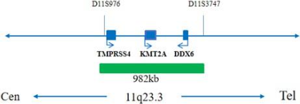
The kit consists of KMT2A/MLLT3 dual color probe as shown in Table 1.

Storage conditions & Validity
Keep sealed away from light at -20oC±5oC. The product is valid for 12 months.Avoid unnecessary repeated freezing and thawing thatshould not exceed 10 times. After opening, within 24 hours for short-term preservation, keep sealed at 2-8oC in dark. For long-term preservation after opening, keep the lid sealed at -20oC±5oC away from light. The kit is transported below 0oC.
Applicable Instruments
Fluorescence microscopy imaging systems, including fluorescence microscopy and filter sets suitable for DAPI (367/452), Green (495/517),and Orange (547/565).
Sample Requirements
1. Applicable specimen type: unfixed fresh bone marrow specimen (stored at 2-8°C for no more than 24 hours).
2. Sample collection take 1-3ml of bone marrow cells anticoagulated with heparin sodium.
3. Sample preservation: after fixation, the cell suspension shall be stored at -20±5°C for no more than 12 months; The prepared cell slides can be stored at -20±5°C for no more than 1 month. When the sample storage temperature is too high or too low, or the cell suspension is volatilized excessively or polluted during storage, the sample will not be used for detection.
Related Reagents
The following reagents are required for the experiment but not provided in this kit.
- 20×SSC, pH 5.3±0.2
Weigh 176g of sodium chloride and 88g of sodium citrate, dissolve in 800mL of deionized water, adjust the pH to 5.3±0.2 at room temperature, and complete to 1 L with deionized water. High-pressure steam sterilization, stored at 2-8oC,the solution shelf life is of 6 months. Discard if the reagent appears cloudy (turbid) or contaminated.
- 2×SSC, pH 7.0±0.2
Take 100mL of the above 20xSSC, dilute with 800mL deionized water, mix, adjust the pH to 7.0±0.2 at room temperature, complete to 1L with deionized water, stored at 2-8oC, the shelf life is of 6 months. Discard if the reagent appears cloudy (turbid) or contaminated.
- Ethanol Solution: 70% ethanol, 85% ethanol
Dilute 700ml, 850ml of ethanol with deionized water to 1L. The shelf life is of 6 months. Discard if the reagent appears cloudy (turbid) or contaminated.
- 0.3% NP-40/0.4xSSC solution, pH 7.0-7.5
Take 0.6mL NP-40 and 4mL 20×SSC, add 150mL deionized water, mix, adjust the pH to 7.0-7.5 at room temperature, with deionized water complete to a volume of 200mL. Stored at 2-8oC, the shelf life is of 6 months. Discard if the reagent appears cloudy (turbid) or contaminated.
- Fixation solution (methanol: glacial acetic acid = 3:1)
Prepare a ready to use fixation solution by mixing thoroughly 30ml of methanol and 10ml of glacial acetic acid.
- 0.075M KCl solution
Weigh 2.8g of potassium chloride, dissolve in 400mL of deionized water and complete to 500mL with deionized water. Stored at room temperature, the solution shelf life is of 6 months. Discard if the reagent appears cloudy (turbid) or contaminated.
- Diamidinyl phenylindole (DAPI) counterstain
Use commercially available anti-quenching DAPI counterstain.
Instructions
- Sample collection and slides preparation
- Sample collection: Take 1-3mL of anticoagulated bone marrow cell samples.
- Cell harvesting: Place the anticoagulated peripheral blood in a 15 mL centrifuge tube, centrifuge at 500g for 5 min, carefully discard the supernatant, and resuspend about 500μL of the residue.
- Cell washing: Add 5 mL of 1×PBS buffer, mix and resuspend the cell pellet, centrifuge at 500g for 5 min, carefully discard the supernatant,and resuspend the cells with about 500μL of the residue; repeat 1 time.
- Cells hypotonicty: Add 10mL of hypotonic solution pre-warmed to 37oC and place in an water bath at 37oC for 15-20min.
- Cells pre-fixation: Pre-fix the cells by adding 1mL (10% by volume) of fixative solution to the cell suspension after the completion of hypotonic osmosis. Gently pipette, mix and centrifuge for 5 min at 500g, discard the supernatant, and resuspend about 500μL of the residue.
- Cell fixation: Slowly add 10mL of fixative solution to the cell suspension at room temperature for 10 min, centrifuge at 500g for 5 min, and resuspend the cells with about 500μL of the residue; repeat once (the cells may be fixed several times until the cells pellet is washed and cleaned).
- Cell suspension preparation: Pipet the supernatant and add the appropriate amount of fixative solution to prepare the appropriate cell suspension concentration.
- Slides preparation: Pipet 3-5μl of cell suspension drop onto the slides, put at 56oC for 30min.
- Slide pretreatment procedure
- At room temperature, rinse the glass slides twice with SSC (pH 7.0) solution for 5min each time.
- Place the glass slides in 70% ethanol, 85% ethanol and 100% ethanol and dry for 2 minutes.
- Denaturation and Hybridization
The following operations should be performed in a darkroom.
- Take out the probe put at room temperature for 5min. Mix and centrifuge briefly. Take 10μl droplet in the cell and drop in the hybridization zone, immediately cover 22mmx22mm glass slide area; spread evenly without bubbles the probe under the glass slide covered area and seal edges with rubber (edge sealing must be thorough to prevent dry film from affecting the test results during hybridization).
- Place the glass slides in the hybridization instrument, denature at 88°C for 2 minutes (the hybridizer should be preheated to 88oC) and hybridize at 45°C for 2 to 16 hours.
- Washing
The following operations should be performed in a darkroom.
- Take out the hybridized glass slides, remove the rubber on the coverslip and immediately immerse the slides in a 2xSSC solution for 5 seconds and remove the coverslip.
- Place the slides in a 2×SSC at room temperature for 1 min.
- Take out the slides and immerse in a preheated at 68°C 0.3% NP-40/0.4xSSC solution and wash for 2min.
- Remove the slides and immerse in a 37°C preheated deionized water, wash for 1min and dry the slides naturally in the dark.
- Counterstaining
The following operations should be performed in a darkroom
10μL DAPI compound dye is dropped in the hybridization area of the glass slide and immediately covered. The suitable filter is selected for glass slide observation under the fluorescence microscope.
- FISH results observation
Place the counterstained film under the fluorescence microscope, and first put it under the low-power objective lens (10x) confirm the cell area under the microscope; Go to 40x under the objective lens, find a position where the cells are evenly distributed; Then in the high-power objective (100x) the FISH results of nuclei are observed.
Interpretation of common signal types
MLLT3 gene signal
KMT2A gene signal
Negative: 2 orange 2 green
Positive: 1 orange 1 green 2 fusion
Precautions
1 .The results of this kit will be affected by various factors of the sample itself, but also limited by hybridization temperature and time,operating environment and the limitations of current molecular biology technology, which may lead to wrong results.
2.Users must understand the potential errors and accuracy limitations that may exist in the detection process.
3.All chemicals are potentially dangerous. Avoid direct contact. Used kits are clinical waste and should be properly disposed off.
Manual Approval date & Revision date
V1. 0: Approval date: April 01, 2019.
V1. 2: Revision date: December 7, 2021.