Product Cat. No.: GBS-118

For Research Use Only

RREB1 gene amplification probe reagent. Instructions Manual

Product Name

RREB1 gene amplification probe reagent..

Package Specifications

10 Tests/box

Product Introduction

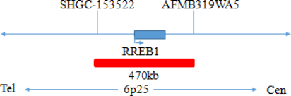
This kit uses orange fluorescein to label RREB1 orange probe and green fluorescein to label CEP6 probes. RREB1/CEP6 dual color probe can be combined with the target detection site by in situ hybridization.

Product Content

This kit consists of RREB1/CEP6 dual color probe, as shown in Table 1.

Table 1: Kit composition

Component name Specifications Quantity Main components
RREB1/CEP6 dual color probe 100μL/Tube 1 RREB1 orange probe ; CEP6 green probe

Storage conditions & Validity]

Keep sealed away from light at -20oC±5oC. The product is valid for 12 months. Avoid unnecessary repeated freezing and thawing thatshould not exceed 10 times. After opening, within 24 hours for short-term preservation, keep sealed at 2-8oC in dark. For long-term preservation after opening, keep the lid sealed at -20oC±5oC away from light. The kit is transported under 0℃.

Applicable Instruments

Fluorescence microscopy imaging systems, including fluorescence microscopy and filter sets suitable for DAPI (367/452), Green (495/517), and Orange (547/565).

Sample requirements

  1. Applicable specimen types: Paraffin-embedded specimensfrom surgical excision or biopsy.
  2. The tissue should be fixed with 4% neutral formaldehyde solution within 1 hour after isolation. After tissue fixation, it is routinely dehydrated and embedded in paraffin.

Test method

  1. Slides pretreatment process
    Baking: Slides heating at 80oC for 30min or 65oC for 2h or overnight.
    Dewaxing: According to the customer laboratory protocol (Commonly with Xylene for 15min).
    Hydration: Take out the slides and put them respectively into 100%, 85% and 70% EtOH at room temperature for 3 minutes each. Take out the slides, and immerse them in deionized water for 3 minutes. Remove the excess of water on the slides by air-drying.
    Permeation: Immerse the slides in deionized water at 100oC and boil continuously for 20-40 minutes (Conventional 20min). Remove the excess of water on the slides by air-drying.
    Digestion: Protease enzymic digestion at 37°C for 10-40 minutes. Mix the protease work buffer (50mmol HCl) and the 10x protease solution (Pepsin concentration 5%) in a proportion of 9:1 to prepare the enzymatic digestion solution.
    Washing: Wash with 2xSSC at room temperature for 5 minutes.
    Dehydration: Take out the slides and dehydrate in 70%, 85%, and 100% gradient ethanol at room temperature for 2 minutes each time. Remove the excess of EtOH solution on the slides by air-drying.
  2.  Denaturation and Hybridization
    The following operations need to be carried out in the darkroom.

    1. Take out the probe, let it stand at room temperature for 5min,turn it upside down with force, fully mix the probe, and then centrifuge briefly (vortex instrument oscillation is prohibited), take 10μL was dropped on the hybridization area of cell drops and immediately covered with 22mm×22mm cover glass, the probe shall be evenly expanded under the cover glass without bubbles, and the edge shall be sealed with rubber glue (the edge must be completely sealed to prevent the dry piece from affecting the test results during hybridization).
    2. Put the tissue sections on the hybridizer, CO denature at 85℃ for 5min (the hybridizer should be preheated to 85℃ in advance), and hybridize at 42℃ for 2-16h.
  3.  Washing
    The following operations need to be carried out in the darkroom.

    1. Carefully remove the sealing glue around the cover glass with tweezers to avoid sticking or moving the cover glass, immerse the sample in 2xSSC for about 5S, take it out, gently push a corner of the cover glassto the edge of the slide with tweezers, and gently remove the cover glass with tweezers.
    2. Place the sample at 2xSSC room temperature for 1 min.
    3. Take out the slides and immerse in a preheated at 68°C 0.3% NP-40/0.4xSSC (Preparation of 0.3% NP-40/0.4xSSC: For 1L preparation, take 3mL NP-40 and 20mL 20xSSC, dissolve fully, mix well, and use 1M NaOH to adjust the pH to 7.2) solution and wash for 2min.
    4. Take out the sample and immerse it in deionized water preheated at 37oC in advance for 1min; dry it naturally in the dark environment.
  4. Counterstaining
    The following operations should be performed in a darkroom
    10μl DAPI compound dye is dropped in the hybridization area of the glass slide and immediately covered. The suitable filter is selected for glass slide observation under the fluorescence microscope.
  5. FISH results observation
    lace the counterstained film under the fluorescence microscope, and first put it under the low-power objective lens (10x) confirm the cell area under the microscope; Go to 40x under the objective lens, find a position where the cells are evenly distributed; Then in the highpower objective (100x) the FISH results of nuclei are observed.

Common Signal Type Interpretation

RREB1 gene signal
CEP6 signal
dots

Negative : 2 orange 2 green

dots

Positive : n orange 2 green (n<=1)

Precautions

  1. The results of this kit will be affected by various factors of the sample itself, but also limited by hybridization temperature and time, operating environment and the limitations of current molecular biology technology, which may lead to wrong results.
  2. Users must understand the potential errors and accuracy limitations that may exist in the detection process.
  3. All chemicals are potentially dangerous. Avoid direct contact. Used kits are clinical waste and should be properly disposed off.

Manual Approval date & Revision date

V1. 0: Approval date: November 01, 2022